
Figure 3.
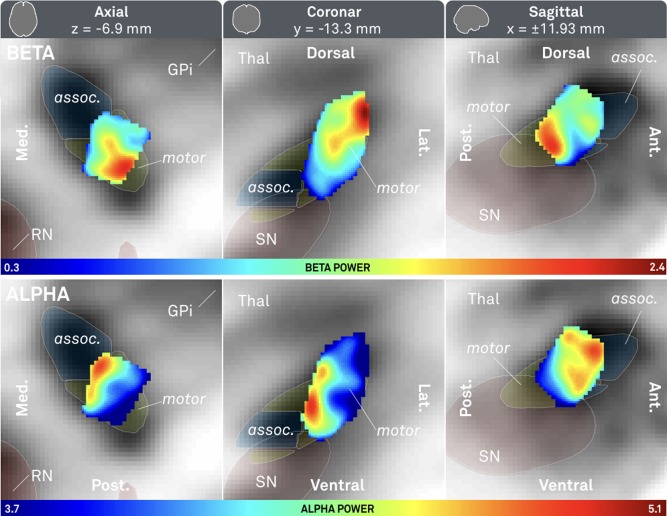
Probabilistic atlas of beta (top row) and alpha (bottom row) power recorded from 309 contact pairs in 54 patients. Axial (left), coronal (mid) and saggital (right) cuts. Associative (assoc.) and sensorimotor (motor) parts of subthalamic nucleus defined by Accolla STN atlas [Accolla et al., 2014]. Red nucleus (RN), internal part of the pallidum (GPi) and substantia nigra defined by ATAG atlas [Keuken et al., 2014]. Note that the beta peak is located more dorsolateral (x = ±14.4, y = −13.2, z = −4.8) whereas the alpha peak is located ventromedial (x = ±13.5, y = −10.3, z = −2.9).