
Figure 6.
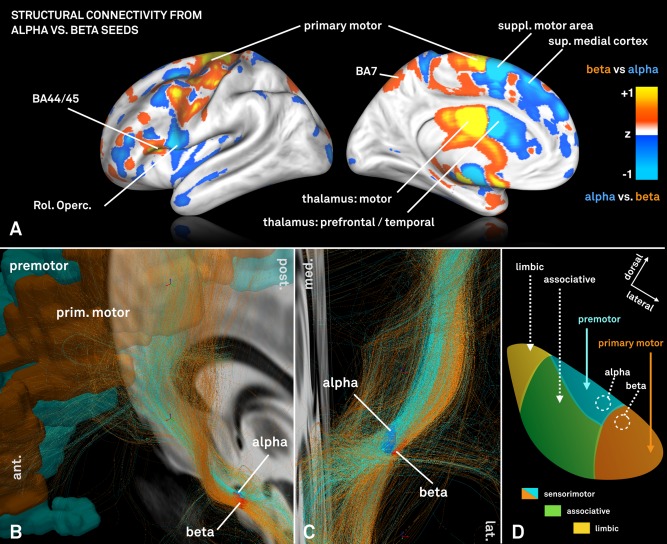
A: Visualization of normalized connectivity profiles when seeding from the electrophysiologically defined alpha versus beta peak on a HCP group connectome. The beta peak is predominantly more connected to primary motor cortices and motor functional zones of the thalamus as defined by the Oxford thalamic connectivity atlas [Behrens et al., 2003]. In contrast, the alpha seed is relatively stronger connected to supplementary motor and other premotor areas such as post. and sup. medial cortices. B + C: Fibertracts traversing through the same electrophysiologically defined alpha and beta seeds within the STN. Two distinct fiber bundles with a different twist angle can be outlined and fibers that traverse through the alpha seed again primarily originate from premotor areas of the cortex whereas fibers selected by the beta seed from primary motor area. D: Schematic representation of STN connectivity that in comparison to Figure 4 further parcellates the sensorimotor STN into areas predominantly receiving input from primary motor (red) vs. premotor (cyan) cortices informed by non‐human primate research [panel adapted from Mathai and Smith, 2011].