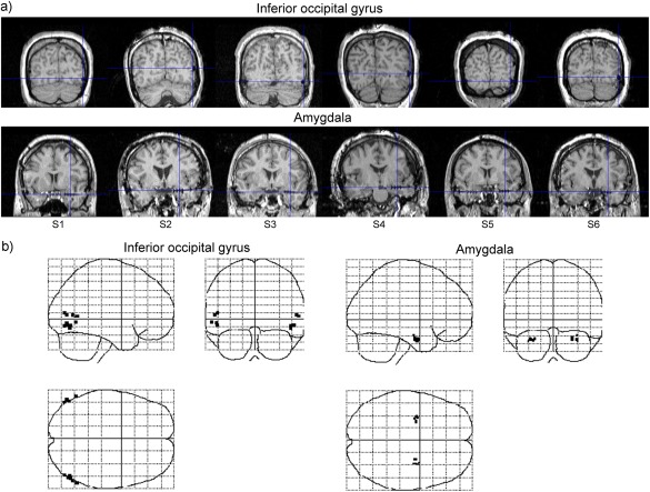
Figure 1.

(a) Anatomical magnetic resonance images of participants. Blue cross‐hairs indicate electrode locations in the inferior occipital gyrus and amygdala in the right hemispheres. Numbers with the prefix “S” indicate the participants' identifications. (b) Glass brain projections of the electrode coordinates (black points) in the Montreal Neurological Institute space.
