
Figure 2.
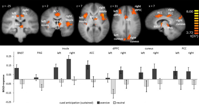
Sustained fear: During the entire anticipation interval, participants showed greater activation in the right bed nucleus of stria terminalis (BNST), periaquaductal gray (PAG), left and right insula, anterior cingulate cortex (ACC), left and right dlPFC, cuneus, and posterior cingulate cortex (PCC) during the anticipation of aversive vs. neutral sounds. Statistical parametric maps are overlaid on an averaged T1 scan. The graph below displays parameter estimates per condition (mean ± standard error for the maximally activated voxel). [Color figure can be viewed in the online issue, which is available at http://wileyonlinelibrary.com.]