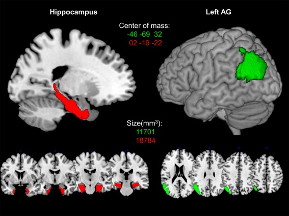
Figure 1.

The red color indicates the bilateral hippocampus ROI, comprising areas CA1, CA2, CA3, subiculum, and entorhinal cortex. The green color shows the left AG ROI, comprising the areas PGa and PGp. ROIs are presented on a 3D rendered surface and coronal slices for bilateral hippocampus and axial slices for left AG, respectively. MNI coordinates of the center of mass and size of every ROI in mm3 is denoted in the corresponding color.
