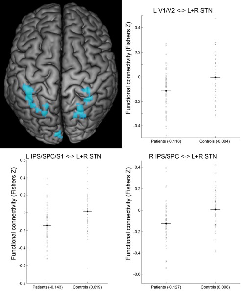
Figure 3.

Enhanced negative coupling with STN in Parkinson patients. Clusters showing a significant PD‐related enhancement of negative coupling (i.e., functional connectivity in patients < controls and functional connectivity negative in patients) with the seed region (i.e., bilateral STN) are marked in cold colors. Surface rendering was performed with MRIcron [Rorden and Brett, 2000]. Scatter plots show functional connectivity of STN (Fisher's Z) with left areas 17 + 18 of visual cortex (L V1/V2), left intraparietal sulcus/superior parietal/somatosensory cortex (L IPS/SPC/S1) and right intraparietal sulcus/superior parietal cortex (R IPS/SPC) for patients and controls. A conjunction of left and right STN seeds was used for data analysis. [Color figure can be viewed in the online issue, which is available at http://wileyonlinelibrary.com.]
