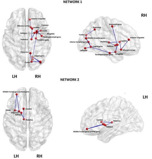
Figure 4.

Networks identified to be significantly different between the de novo PD and control groups using Network‐Based Statistical Analysis. Netwotk1 consisted of 16 edges connecting 16 different regions (corrected P = 0.009). These connections primarily involved key components of the limbic system, basal ganglia and sensorimotor areas confirming the involvement of this regions in the pathophysiology of PD. Network2 consisted of six edges linking six nodes (corrected P = 0.018). These edges connected the left thalamus to both left caudate and the left pallidum, the left pallidum to the left putamen, the left putamen to both the left insula and the left middle frontal gyrus (orbital part), and the left insula to the left middle frontal gyrus (orbital part). LH, left hemisphere; RH, right hemisphere. [Color figure can be viewed at http://wileyonlinelibrary.com]
