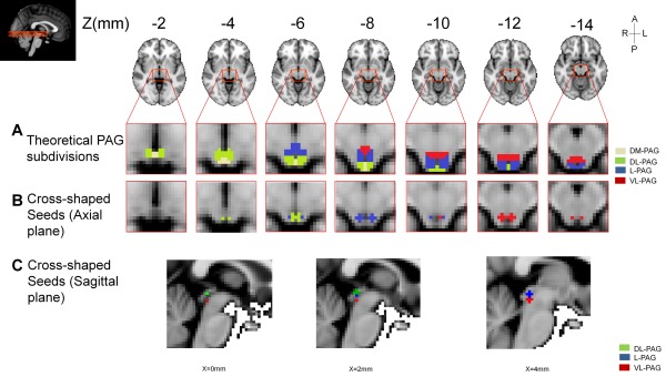
Figure 1.

Theoretical subdivision of the PAG (A) and seeds used in the seed‐to‐whole brain analysis (B and C) shown in 2 mm3 standard space. Seeds were designed as 2‐mm‐radius spherical seeds in 1 mm standard space (cross‐shaped once transformed to 2 mm3 standard space and binarized). 1 mm standard space MNI coordinates: VL‐PAG (red) [±3, −32, −12], L‐PAG (blue) [±4, −31, −8], DL‐PAG (green) [±2, −32, −5]. R: right; L: left; A: anterior; P: posterior. The theoretical subdivision of the PAG is provided to illustrate the uneven distribution of the DL‐, L‐, and VL‐PAG in the inferior part of the PAG compared to rostral part of the PAG, for example, the superior part of the PAG is composed mainly of dorsal PAG (medial and lateral) and the inferior part of the PAG is composed mainly of lateral and ventrolateral PAG, with a smaller portion for the dorsal PAG. Seeds shown on sagittal plane are presented in Supporting Information (Fig. S1). For a complete schematic representation of the PAG, see Bandler and Shipley [1994] and Linnman et al. [2012b].
