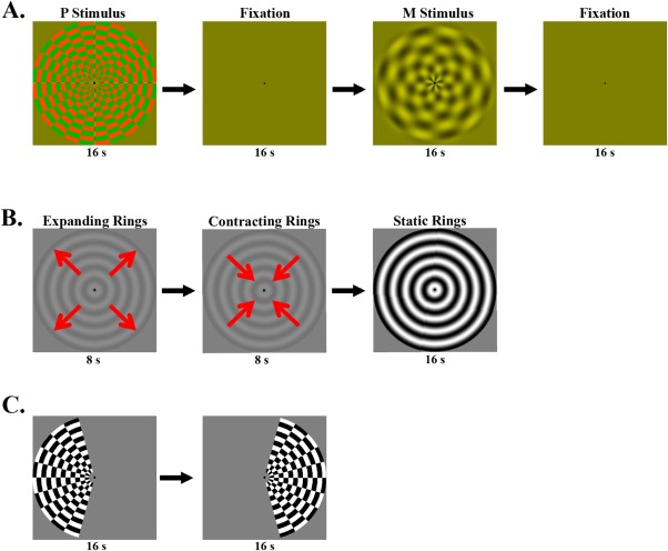
Figure 1.

(A) Stimuli for the M and P localizer. The M and P stimuli were presented in blocks of 16 s duration, alternating with 16 s fixation. (B) Stimuli for the MT localizer. Each stimulation cycle started with a low contrast ring expanding for 8 s, and then contracting for 8 s, followed by a static ring of high contrast presented for 16 s duration. C. Stimuli for the LGN localizer. The hemifield checkerboards alternatively presented every 16 s between the left and right visual field. [Color figure can be viewed in the online issue, which is available at http://wileyonlinelibrary.com.]
