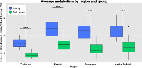
Figure 3.

PET glucose uptake in DMN regions. Standardized uptake value following partial volume correction (PVC) in the default mode network regions (average of standardized uptake values of left and right hemisphere) for healthy controls and brain injured patients. Brain injured patients show a decreased standardized uptake value compared with controls in all default mode network regions. *** = P < 0.001. [Color figure can be viewed at http://wileyonlinelibrary.com.]
