
Figure 4.
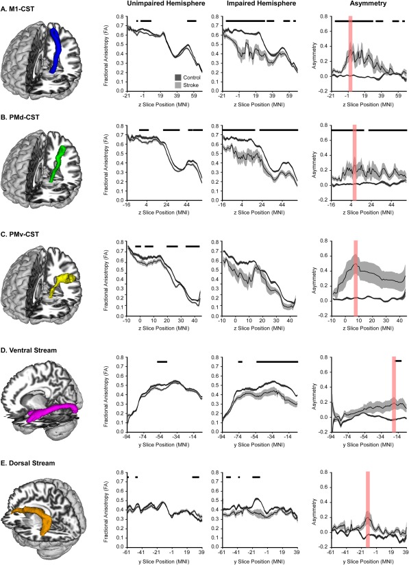
Probabilistic Tractography in Motor and Visual Tracts. FA profiles of all motor and visual tracts. The mean FA for each group is displayed with a black line, and the dark gray (control) and light gray (stroke) shaded areas represent ± SEM for each group. Comparisons were made in the unimpaired hemisphere and impaired hemisphere, with FDR corrected P < 0.05 represented with horizontal black lines in each plot. Asymmetry of each slice within each tract was also calculated, and is displayed in the final column. The slice with the highest asymmetry is highlighted in red, and represents the region used to extract data for the multiple regression analyses.