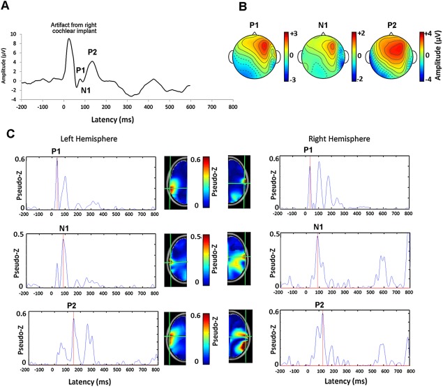
Figure 1.

A. Example of a cortical evoked waveform at Cz recorded from one adolescent with 15.95 years of CI experience in the right ear indicates a mature response with peaks P 1, N 1 and P 2. The electrical artifact from the right CI preceding this response is clear. B. Head topographies of potential distribution for each peak (common averaged reference) of the mature cortical response are shown. C. The activity underlying the mature peaks in 63,646 brain voxels are shown relative to the noise floor using the pseudo‐Z. The virtual channel waveforms corresponding to the voxel with the strongest activity (red hotspots) are shown for the left and right hemispheres for each peak. Source locations are marked by the green cross hairs on the axial view of the age‐appropriate topographic head model derived from the MNI MRI Library. The virtual channel data reveals that the CI artifact has been suppressed in early latencies by the beamforming procedure and that the strongest dipole underlying each peak of the mature response occurs in areas of the left and right auditory cortices, in line with the latency of the cortical wave peaks.
