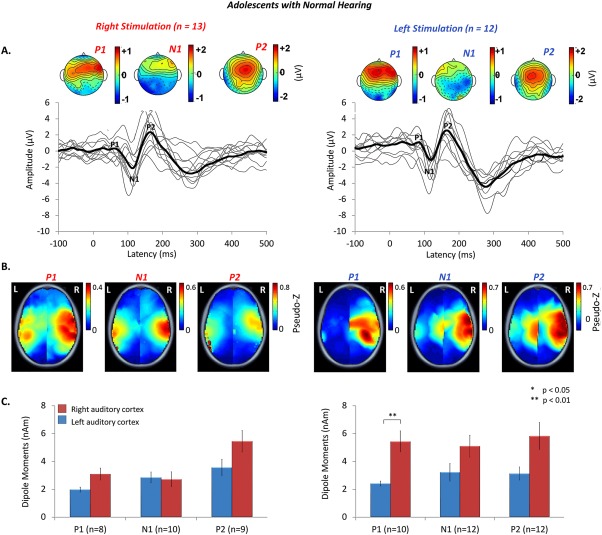
Figure 2.

A. Auditory evoked cortical responses were stimulated from the right (n = 13) and left (n = 12) ears of adolescents with normal hearing, aged 15.9 ± 6.4 years. Individual responses from a midline cephalic electrode (C z) (thin grey lines), and grand mean responses (thick black lines) show three peaks (P 1, N 1, P 2), characteristic of mature responses. Topographic maps indicate similarly distributed scalp potentials (red = positive potentials, blue = negative potentials) for the mature peaks in both ears (common averaged reference). B. Mean peak dipole activity reveals stronger activity, shown by hotspots of dark red, in the right hemisphere, particularly for left ear stimulation. C. Mean [± 1 standard error (SE)] dipole moment strength is plotted for the left (blue bars) and right (red bars) auditory cortices for all cortical peaks. Consistent with the topographic plots in B. peak dipole activity is stronger in the right than left auditory cortex for stimulation of both ears, particularly for left ear stimulation.
