
Figure 1.
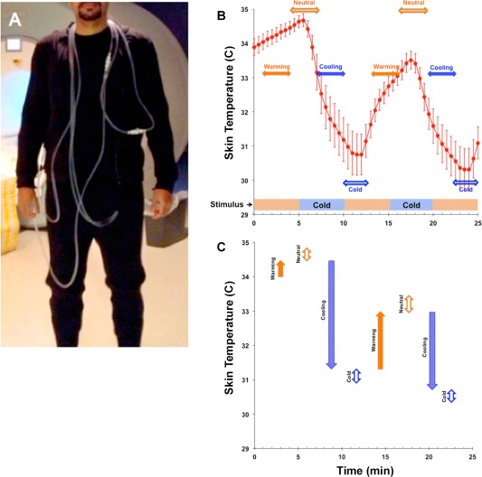
(A) Subject dressed in the tube suit covering the arms to the wrists, the legs to the ankles and the torso. (B) The bar at the base of the graph depicts the stimulus (study paradigm) consisting of two 5‐min cooling periods (blue/dark) interspersed between neutral temperature background (orange/light), resulting in average skin temperature oscillations (error bars ± s.d.). From the temperature curve, we derived two classes of epoch windows (horizontal arrows). The filled arrows depict temporal windows characterized by warming (orange/light) or cooling (blue/dark). Complementary temporal windows (open arrows) assessed fMRI responses for neutral (orange/light) or cold (blue/dark) periods. These periods were characterized by different ranges of skin temperature. (C) The vertical arrows depict the range (and direction of change) of skin temperature during warming or cooling, and neutral or cold temporal windows. Color/Shading conventions are maintained from (B). The figure clearly indicates that periods of warming and cooling were associated with more dynamic changes in skin temperature than periods of neutral or cold. [Color figure can be viewed in the online issue, which is available at http://wileyonlinelibrary.com.]