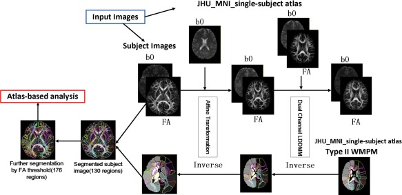
Figure 2.

Schematic diagram of dual‐contrast LDDMM Atlas‐based analysis pipeline. FA and b0 maps of each participant were first transformed linearly and then non‐linearly to “JHU_MNI_single‐subject” atlas. Next, the inverse transformation was applied to the pre‐segmented WM atlas, thus enabling automated segmentation of the original images into 130 subregions. The cortex and the associated SWM were further divided using a threshold (FA > 0.25) for each subject, resulting in a total of 176 subregions. [Color figure can be viewed in the online issue, which is available at http://wileyonlinelibrary.com.]
