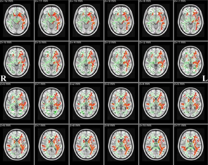
Figure 3.

FA skeleton clusters at P < 0.05 (family‐wise error corrected for multiple comparisons). We overlay the FA skeleton (green) on axial slices of the standard MNI_T1_1mm template. Red‐Yellow voxels represent regions with significantly decreased FA values in Tourette syndrome patients relative to healthy controls. The white‐matter microstructure is altered in the body of corpus callosum, forceps major, left superior longitudinal fasciculus, ATR/superior cerebellar peduncle, right superior longitudinal fasciculus (temporal part), inferior longitudinal fasciculus, inferior fronto‐occipital fasciculus and corticospinal tract. [Color figure can be viewed in the online issue, which is available at http://wileyonlinelibrary.com.]
