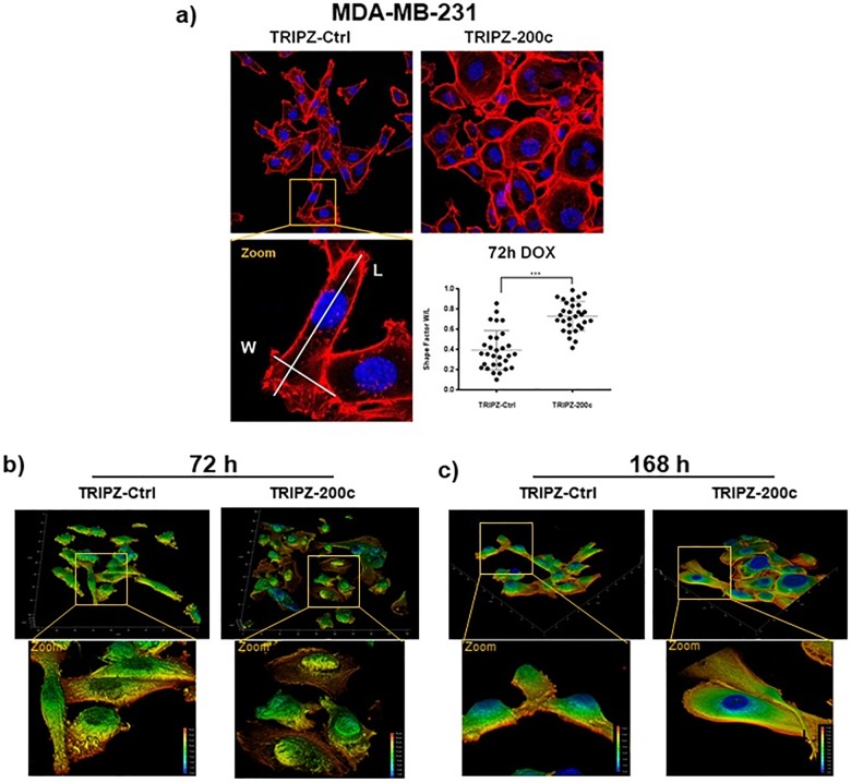
Fig 3. Overexpression of miR-200c induced fast morphological changes in MDA-MB-231 cells.
(a) Fluorescence staining of the actin cytoskeleton by phalloidin (red) and nuclei (blue) in MDA-MB-231 decreased spindle-like phenotype after induction miR-200c, as shown by significant changes in the shape factors (N = 30; error bars are SD; *** p > 0.001). (b, c) Renderings of z-stacked immunofluorescence images of MDA-MB-231 acquired by confocal imaging showed decreased mesenchymal shape in 3D after induction of miR-200c for (b) 72 h as well as (c) further increased effects after 168 h compared to included controls.

