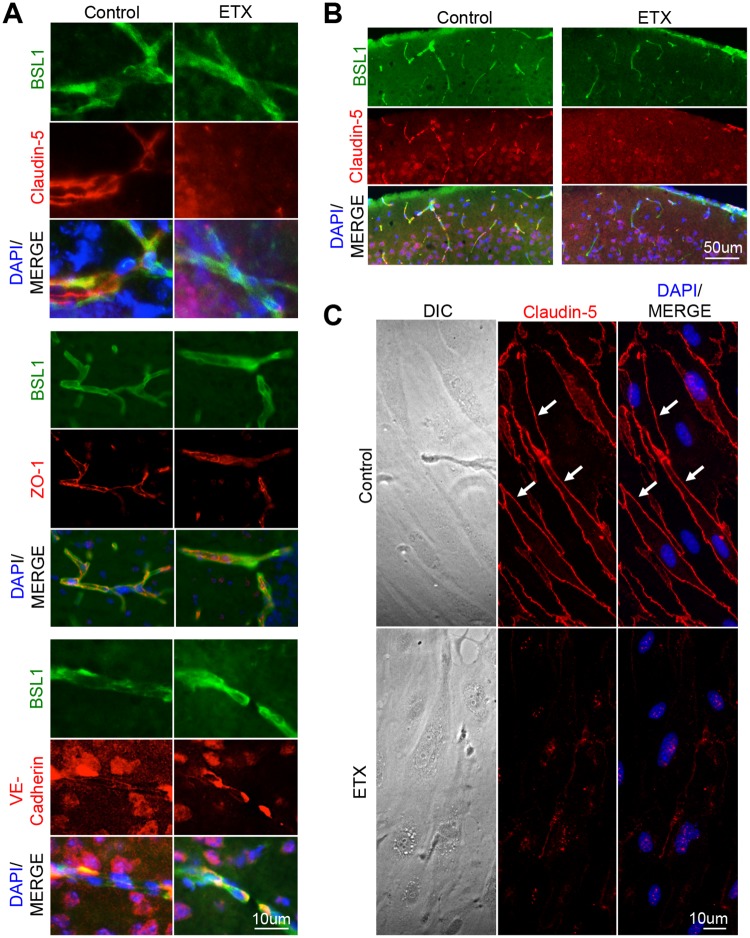
Fig 7. ETX treatment causes a down regulation in claudin-5 staining.
Mice were treated with 5ng of ETX per gram body weight for one hour and then perfused with PBS. Saline treated mice were used as controls. (A) Cyrsosections were evaluated for tight junction markers including claudin-5, ZO-1, and VE-cadherin. FITC-BSL1 (green) was used to visualize vasculature. (B) Extensive reduction in claudin-5 staining was also observed in ETX treated animals compared to controls. Sagittal sections of cortical matter. (C) Down regulation of claudin-5 was also confirmed in-vitro. Primary mouse BEC were treated with or without 50nM ETX for two hours and then stained for claudin-5. Under control conditions, claudin-5 is found at cell-cell junctions (white arrows). When treated with ETX, claudin-5 treatment significantly decreases and becomes perinuclear.

