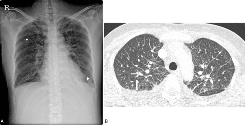
Figure 2.

Chest radiography and CT of a patient with scrub typhus. A. Chest radiograph shows diffuse and mild interstitial thickening ( ) in the bilateral lungs. A small amount of bilateral pleural effusion (▾) is noted. B. Axial chest CT shows smooth interlobular septal line thickening (
 ), axial interstitial thickening, and bilateral pleural effusion (
 ). CT = computed tomography.
