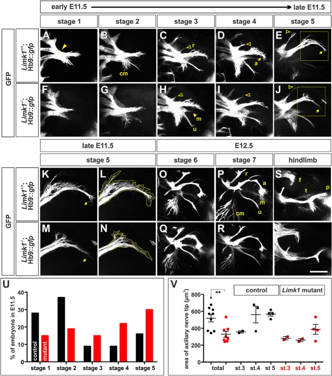
Figure 3.
Motor axon branching is advanced in E11.5 Limk1 mutant embryos compared with controls. A–T, Using whole-mount limb preparations, we established the timeline (Stages 1–7) over which motor nerves project into either the forelimb (A–R) or hindlimb (S, T; and data not shown) of E11.5 (A–N) to E12.5 (O–T) Hb9::gfp transgenic embryos, either alone (control, A–E, K, L, O, P, S) or in combination with the Limk1 mutation (mutant, F–J, M, N, Q, R, T). The pattern by which the GFP+ branches segregated was highly reproducible between embryos; however, the ventral nerve branches, especially the median (m) nerve, often extended out of the plane most easily visualized by confocal microscopy. A, At Stage 1, the earliest stage observed in E11.5 embryos, the MNs in the brachial lateral motor column have projected into and past the brachial plexus (arrowhead) and have just divided to innervate the dorsal and ventral forelimb. B, Stage 2 is marked by the ventral branch segregating into the cutaneous maximus (cm), ulnar (u), and median nerves (see also H). C, Stage 3 is marked by the emergence of the radial (r) nerve (open arrowhead). D, E, Stages 4 and 5 are distinguished by the extent of growth of the radial nerve (open arrowheads) and the increasing branching of the cutaneous maximus nerve. In the later stages, the axillary (a) nerve starts (Stage 4) and then completes (Stage 5) a highly reproducible turn (E, J, arrows). F–J, Limk1 mutant motor nerves branch with a similar pattern to those in control littermates. However, the GFP+ Limk1−/− axons in the axillary nerve tip appear to be less exploratory as they reach the turning point in stages 4 and 5 (J, box). K–N, High-magnification images of the control (K, L) or mutant (M, N) axillary nerve tip shown in the boxed region of E and J, respectively. L, N, Outline of the area of the tip used for quantification in V. O, P, Stages 6 and 7, the stages in our timeline occurring in E12.5 embryos, are marked by the increasing extent of the cutaneous maximus nerve and length of the axillary nerve. Both the radial and axillary nerves start to bifurcate in Stage 6; this bifurcation is pronounced at Stage 7. Q, R, By E12.5, the branching pattern of Stage 6 and Stage 7 GFP+ Limk1 mutant motor nerves is indistinguishable from controls. S, T, By E12.5 in the hindlimb, GFP+ motor nerves have projected from the lumbar plexus into three branches: the femoral (f), dorsal peroneal (p), and ventral tibial (t) nerves. There was no distinguishable difference between the branching patterns in the control (S; n = 10) and Limk1 mutant (T; n = 3) hindlimbs. U, E11.5 embryos are not evenly distributed between the five stages. Whereas control forelimbs (n = 43 limbs) are more likely to be either Stage 1 or Stage 2, mutant forelimbs (n = 27 limbs) are more likely (probability that the distribution of control stages is similar to the mutants: p < 0.0025, Fisher's exact test) to be either Stage 4 or Stage 5, strongly suggesting that the extension of Limk1−/− motor nerves is more advanced in development than controls. V, The total area of the axillary nerve tip is ∼35% smaller in Stage 3–5 Limk1 mutant forelimbs (n = 8 embryos) compared with Stage 3–5 control littermates (n = 9 embryos, probability of similar, p < 0.0022 Student's t test). Interestingly, the area of control nerve tips expands rapidly between Stage 3 and Stage 4; this expansion is observed in Limk1 mutant nerve tips, but not until Stage 5. This result suggests the response of the accelerated Limk1−/− axons to the surrounding environment is delayed until the appropriate time in development. Scale bars: A–J, 250 μm; K–N, 150 μm; O–T, 500 μm. Probability of similarity, **p < 0.005, Student's t-test.

