Figure 4.
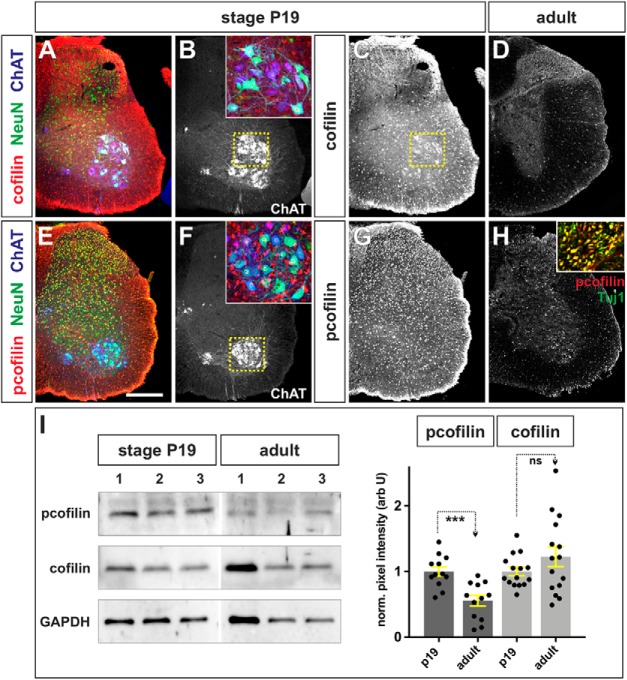
Cofilin is largely inactive in the adult spinal cord. A–H, Limb level sections of P19 (A–C, E–G) or adult (D, H) spinal cords were labeled with antibodies against total cofilin (red, A, C, D), phosphorylated (p)-cofilin (red, E, G, H), NeuN (green, neuronal nuclei; A, E), ChAT (blue, mature MNs; A, B, E, F), and Tuj1 (green, neuronal processes; H, inset). A–C, Cofilin is present at high levels throughout the P19 postnatal spinal cord. In this lumbar section, cofilin is present at high levels in a population of ventral NeuN+ ChAT+ neurons, thought to be α MNs (magnified B, inset). D, Cofilin continues to be expressed in the adult spinal cord, albeit at significantly lower levels, particularly in the white matter. E–H, Cofilin is highly phosphorylated at both neonatal (E–G) and adult stages (H), including in the ventral NeuN+ ChaAT+ MNs (F, inset) and the Tuj1+ neural processes in the white matter (H, inset; and data not shown), suggesting that cofilin is mostly inactive in axons. I, The levels of cofilin and (p)-cofilin were quantified in a Western analysis of P19 and adult ventral spinal cord tissue, using GAPDH levels as a loading control. While cofilin levels are not significantly different (p > 0.17, Student's t test) between the P19 and adult spinal cord (three independent samples shown in I; n = 6 animals analyzed in 5 experiments), there was a ∼50% decrease in (p)-cofilin levels in adult spinal cord (n = 6 animals analyzed in 4 experiments; p < 0.0004, Student's t test) compared with P19 animals. Scale bars, 300 μm. Probability of similarity, ***p < 0.0005, Student's t-test.

