Figure 1.
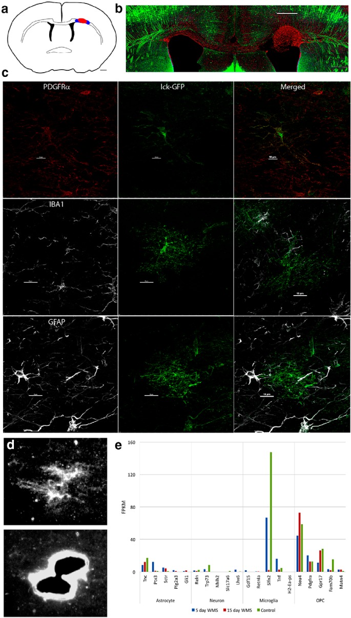
White matter stroke and LCM. a, Schematic of location of white matter stroke (red) in coronal section of mouse brain. Blue areas represent regions of LCM of OPCs. Scale bar, 500 μm. b, White matter stroke in YFP-H mouse in which layer 5 neurons are densely labeled. The red cells are DAPI stains, with pseudocolor to red for visualization. Scale bar, 300 μm. c, The specificity of the viral labeling of OPCs (lck-GFP) in white matter is shown with coimmunohistochemical staining for markers of microglia (IBA1), astrocytes (GFAP), and OPCs (PDGFRα). Scale bar, 50 μm. d, Viral labeling of OPC and then laser dissection of OPC for downstream RNA isolation and sequencing. Lentivirus with membrane-bound GFP (lck-GFP) under the control of the PDGFRα promoter is injected into the white matter adjacent to the stroke (a, blue region), labeling OPCs. These are microdissected in the region of partial damage to white matter, adjacent to the stroke site. e, To assess the OPC specificity of the RNAseq dataset, fragments per kilobase of transcript per million reads mapped (FPKM) data for each of the three treatment groups were compared with the most cell type-specific genes identified in an online database (Zhang et al., 2014). FPKM values for the top 10 most cell type-specific genes of the indicated type cell type are compared with the day 5 and day 15 regenerating OPCs transcriptomes in white matter stroke.

