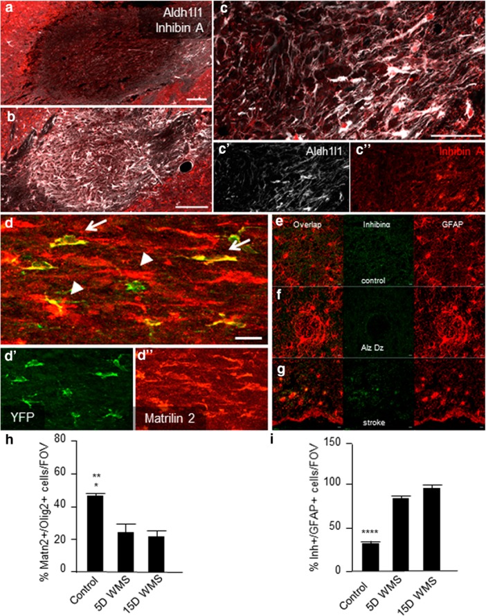
Figure 4.
Astrocyte and OL/OPC Matrillin-2 and Inhibin A signaling. a–c, Inhibin A subunit Inhibinα is used as a marker to compare tissue levels of the growth factor (red), Aldh1l1 (white) labels astrocytes. At 5 d after lesion (a), there is no detectable Inhibinα signal within the stroke, whereas labeling of 15 d stroke sections (b) revealed high levels of Inhibinα+ cells filling the lesion. In higher magnification, at the edge of the stroke. c–c″, The majority of Inhibinα label colocalizes with Aldh1l1+ astrocytes shown in higher magnification. Inhibinα (red) and Aldh1l1 immunohistochemistry 5 d after stroke (a) and 15 d after stroke (b). c, Higher magnification shows colocalization of Aldh1l1 staining with inhibinα. Scale bars: a–c, 100 μm; c′, 20 μm. d, In a NG2-CreERT2/r26YFP mouse, OPC reporter line 5 d after stroke. d′, YFP+ (yellow) OPCs colabel with d″, Matrilin-2 (red) (arrows) in addition to other cell types (arrowheads). Scale bar, 100 μm. e–g, Inhibinα staining in human white matter from control (e), Alzheimer's disease as a reactive astrocyte condition that is not stroke and in white matter stroke (f). Scale bar, 20 μm. h, i, Quantification of percent immunopositive staining for Matrilin-2 and Inhibinα in Olig2+ cells in control and after stroke. *p = 0.0111 versus 5 d white matter stroke. **p = 0.0040 versus 15 d white matter stroke. ****p < 0.0001.

