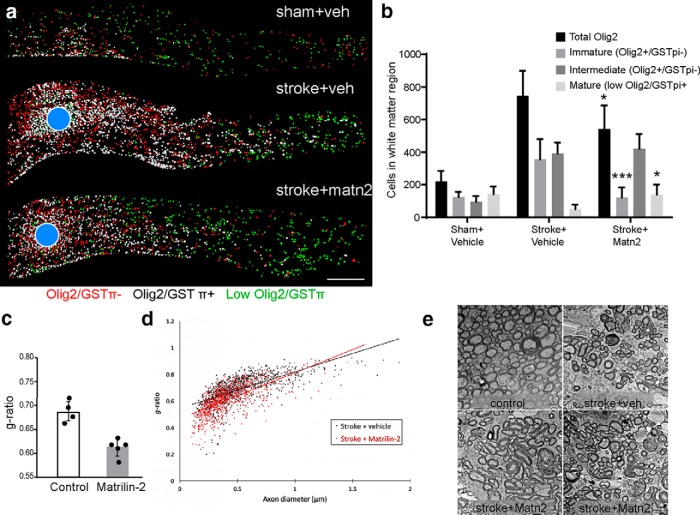
Figure 9.
Matrilin-2 promotes tissue measures of OPC differentiation in vivo. a, Distribution of OLs in the subcortical white matter was mapped 8 weeks after stroke. The number of spots in each category of mature OL (low olig2/GSTπ+), intermediate OL (Olig2+/GSTπ+), and immature OL or OPC (Olig2/GSTπ−) and their distance from the stroke lesion center (blue circles) mapped. Blue circle represents the center of the stroke. b, Quantification of OL lineage cells in control, stroke, and stroke+Matrilin-2 delivery, 8 weeks after stroke. *p < 0.05 versus Stroke+veh. ***p < 0.001 versus Stroke+veh. c, d, G ratio was measured 9 weeks after stroke. The data are the mean g ratio for each animal (n = 5 for each group) ± SD. e, Representative EM images from corpus callosum that were used to calculate g ratios, showing the different conditions in aged white matter, stroke, and two examples of exogenous matrilin-2 delivery.

