. 2019 Oct 31;8:e52654. doi: 10.7554/eLife.52654

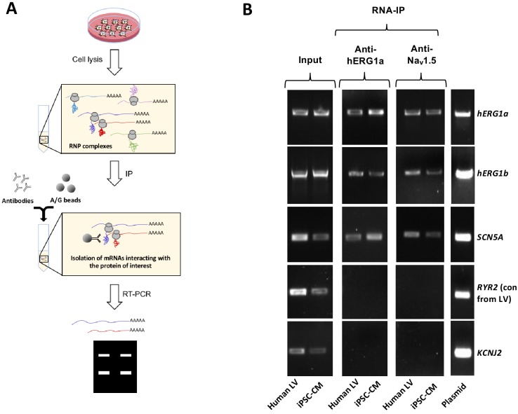
Figure 1. Complex of ion channel transcripts with nascent proteins.

(A) Scheme of the RNA-IP protocol in which channel-specific antibodies are used to pull down nascent proteins and associated transcripts. RNP: ribonucleoprotein. (B) Lanes 1 and 2, RT-PCR products from input lysate of human left ventricle (LV), and iPSC-CM. Lanes 3–16 shows the corresponding RNA-IP’s using an anti-hERG1a or anti-NaV1.5 antibodies; Lane seven shows the control (+) and represents signal amplified from purified plasmid template. Similar results were obtained in at least three independent experiments. (N = 5 for anti-hERG1a and N = 3 for anti-Nav1.5 using human LV and iPSC-CMs).

Figure 1—source data 1. RNA-IP Blots raw data for Figure 1B.
DOI: 10.7554/eLife.52654.004

Figure 1.

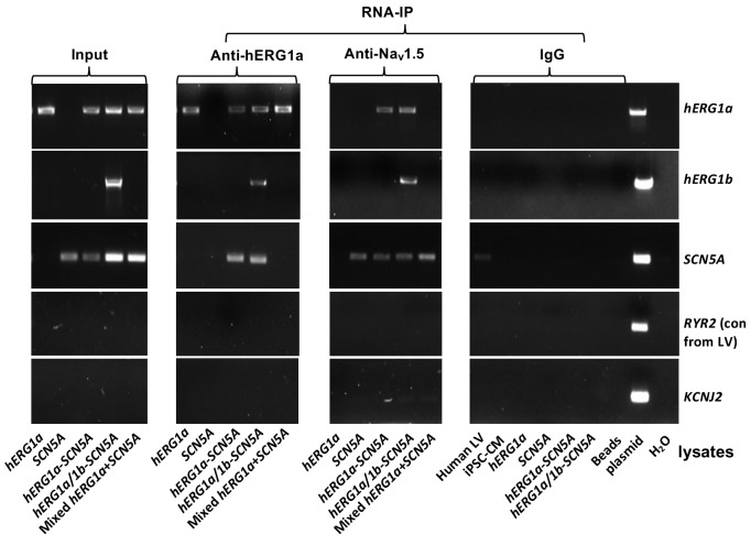
Figure 1—figure supplement 1. Complete RNA-IP from Figure 1.

Figure 1—figure supplement 1.

Lanes 1–6, RT-PCR products from input lysate of human left ventricle (LV), iPSC-CM, and HEK293 cells expressing: hERG1a; SCN5A; hERG1a plus SCN5A; and hERG1a plus hERG1b and SCN5a. Lane seven shows RT-PCR product from lysates independently expressing hERG1a and SCN5A, mixed. Lanes 8–14 shows the corresponding RNA-IP’s using an anti- hERG1a antibody, followed by a bead-only control and H2O control. The next group shows the corresponding RNA-IP’s using the anti-Nav1.5 antibody, followed by a group of IgG controls. H2O and beads lanes show absence of template contamination; control (+) represents signal amplified from purified plasmid template.