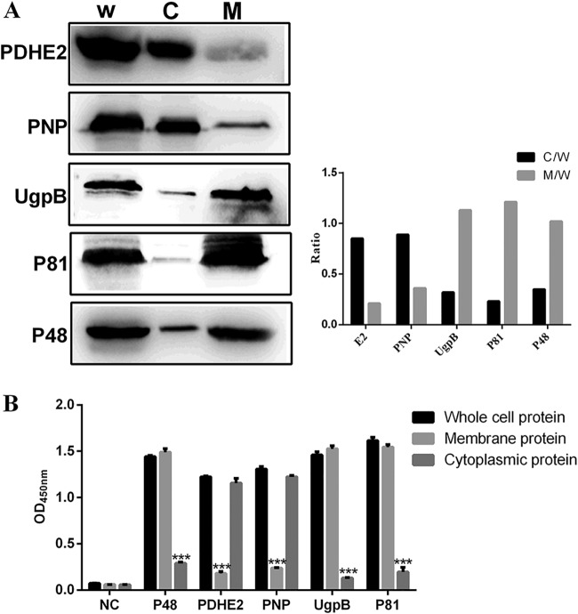
FIG 3.
Subcellular localizations of PDHE2, PNP, UgpB, and P81 in M. bovis. (A) Western blotting with the listed specific antibodies. W, whole-cell protein; C, cytoplasmic protein; M, membrane protein. (Left) Representative blot; (right) graph showing the ratio of the protein amount in the cytoplasmic or membrane fractions to the total protein in whole-cell lysate. (B) For indirect ELISA, 96-well ELISA plates were coated with whole bacterial proteins, membrane proteins, or cytoplasmic proteins. Rabbit anti-PDHE2 serum, rabbit anti-PNP serum, rabbit anti-P81 serum, and rabbit anti-UgpB serum were used as primary antibodies, and HRP-conjugated goat anti-rabbit IgG was used as the secondary antibody. The optical density at 450 nm (OD450) was read. The whole-bacterial protein group was the control group. Data represent means ± SD from 3 independent experiments. Statistical analysis was done using a ratio-paired two-tailed t test and displayed only when significant. ***, P ≤ 0.001.

