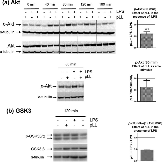
FIG 1.
Exposure to pLL inhibits Akt (a) and GSK3 (b) phosphorylation in LPS-stimulated GM-CSF–BMDCs. GM-CSF–BMDCs were exposed to pLL, LPS, or both stimuli together for the indicated times. Cell lysates were analyzed for phosphorylated and total Akt (a) and for phosphorylated GSK3α and -β and total GSK3β (b). In panel a, the lower Western blot shows that pLL as the sole stimulus does not cause detectable Akt phosphorylation (similar results were obtained for incubation times between 10 and 160 min). The Western blots shown are representative of at least two independent experiments. The graphs show the quotients of p-Akt or p-GSK3α/β values (normalized over loading controls) for cells treated with pLL over cells not exposed to pLL in the presence or absence of LPS, as indicated. The graphs for p-Akt show means and standard deviations (SD) corresponding to six to eight independent experiments. Asterisks (*, P ≤ 0.05; ***, P ≤ 0.001) indicate significance as determined by a one-sample t test for comparison with unity (i.e., no inhibition). The graph for p-GSK3 shows the means and ranges of two independent experiments.

