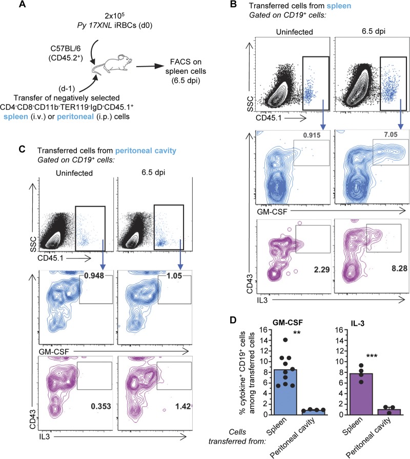
FIG 4.
GM-CSF- and IL-3-producing B cells originate from the spleen of P. yoelii-infected mice. (A) Schematic of experimental design. i.v., intravenous. (B and C) Spleen cells from P. yoelii-infected or uninfected mice were stained for cell surface CD45.1, CD19, and CD43 and intracellular GM-CSF or IL-3. Data show a representative FACS dot plot of GM-CSF+ or IL-3+ cells after gating on transferred CD45.1+ cells isolated from the spleen (B) or peritoneal cavity (C). SSC, side scatter. (D) Average proportion of GM-CSF- or IL-3-producing CD19+ B cells among transferred cells across two independent replicate experiments, with each dot featuring an individual mouse (n = 3 to 10 mice). Graphs shown the average of the results from each experiment along with the SEM. P values are indicated when applicable.

