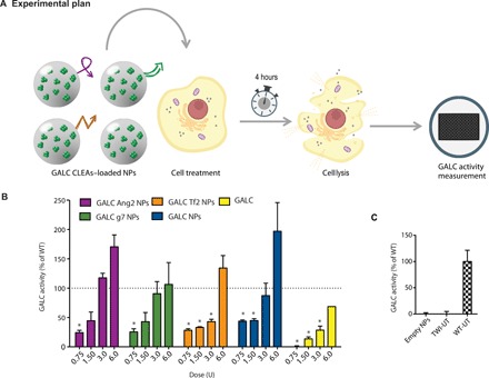
Fig. 3. In vitro ERT: Dose-response experiment.

(A) Graphical summary of the experiment. GALC-deficient primary fibroblasts (derived from TWI mice) were treated with GALC CLEA NPs. After 4 hours, GALC activity was measured on cellular lysates by 6-hexadecanoylamino-4-methylumbelliferyl-β-d-galactopyranoside (HMU-βGal) assay. (B) Dose-response experiment results. Cells were treated with targeted GALC CLEA NPs (GALC Ang2 NPs, GALC g7 NPs, or GALC Tf2 NPs), with nontargeted NPs (GALC CLEA NPs), or with the free rm-GALC (GALC). For every treatment, four doses were tested: 0.75, 1.50, 3.0, and 6.0 U [unit (U) = amount of enzyme that catalyzes 1 nmol of substrate per hour]. Results are expressed in unit per microgram and reported in percentage of the activity of the WT cells [(U/μg) = unit of enzyme per microgram of cell lysate]. *P < 0.05 specific treatment versus WT, one-way analysis of variance (ANOVA) (Dunnett’s multiple-comparisons test), means ± SEM (n = 3). (C) Controls. For control, NPs nonloaded with GALC CLEAs (empty NPs) were also administered to the cells. WT- and TWI-untreated cells activity is also showed. Means ± SEM (n = 3).
