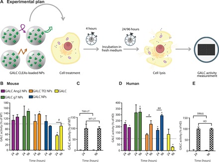
Fig. 4. In vitro: ERT time-response experiment.

(A) Graphical summary of the experiment. GALC-deficient primary murine (from TWI mice) and human (from patients with KD with GALC 30kbΔ) fibroblasts were treated with a single dose [3.0 U; unit (U) = amount of enzyme that catalyzes 1 nmol of substrate/hour] of targeted GALC CLEA NPs (GALC Ang2 NPs, GALC g7 NPs, or GALC Tf2 NPs), nontargeted NPs (GALC CLEA NPs), or with the free rm-GALC (GALC). Four hours later, cells were incubated with fresh medium. GALC activity was measured in the cell lysates 24 or 96 hours later by HMU-βGal assay. (B) NPs mediated ERT in GALC deficient mouse cells. Results are expressed in unit per microgram and reported as percentage of the activity of the WT cells (U/μg = unit of enzyme per microgram of cell lysate). *P < 0.05 specific treatment versus WT, one-way ANOVA (Dunnett’s multiple comparisons test). #P < 0.05 GALC 96 hours versus GALC 24 hours, Student’s t test, means ± SEM (n = 3). (C) Controls. WT and TWI untreated cells activity at 24 and 96 hours. Means ± SEM (n = 3). (D) NPs mediated ERT in GALC deficient human cells. Results are expressed in unit per microgram and reported in percentage of the activity of the healthy (HD, human donor) cells (U/μg = unit of enzyme per microgram of cell lysate). *P < 0.05 specific treatment versus WT, one-way ANOVA (Dunnett’s multiple comparisons test). #P < 0.05 GALC Tf2 NPs 96 hours versus GALC Tf2 NPs 24 hours and ##P < 0.01 GALC NPs 96 hours versus GALC NPs 24 hours, Student’s t test, means ± SEM (n = 3). (E) Controls. HD and 30kbΔ untreated cells activity at 24 and 96 hours. Means ± SEM (n = 3).
