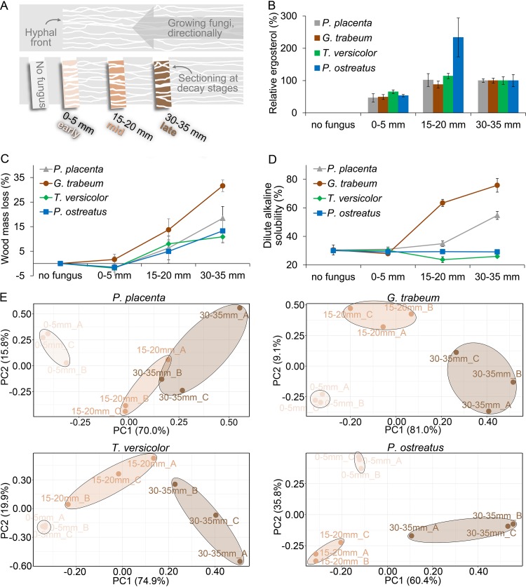
FIG 1.
A fine-scale time series of wood decay using directed fungal growth in wafers. (A) Wood decay stages during brown rot (by P. placenta and G. trabeum) or white rot (by T. versicolor and P. ostreatus) were manually sectioned in aspen wafers, including a section without fungal growth (no fungus) and three sections behind the hyphal front (0 to 5 mm, 15 to 20 mm, and 30 to 35 mm). A total of 24 independent wafers (n = 8, each represented) were sectioned per fungus per experiment, using the hyphal front to benchmark progression. A fungal biomarker, ergosterol (B), is shown relative to late-decay-stage levels (as %) and indicates significant biomass in the 0- to 5-mm “early” section. Wood mass loss (C) and dilute alkaline solubility of wood (D [a sensitive test of early brown rot modifications]) were measured to assess the decay intensity, and neither could distinguish the 0- to 5-mm section from sound wood (ANOVA HSD test, P > 0.1). These results, combined, verified that 0- to 5-mm sections were well-colonized but lacked discernible decay, and principal-component analyses (E) clearly distinguished the whole-genome expression patterns in this “early” section (0 to 5 mm) from those in advanced-decay sections (15 to 20 mm and 30 to 35 mm) for all four test fungi. Three bioreplicates from each section (i.e., A, B, and C) were well clustered, indicating the reproducibility of the RNA-seq results.

