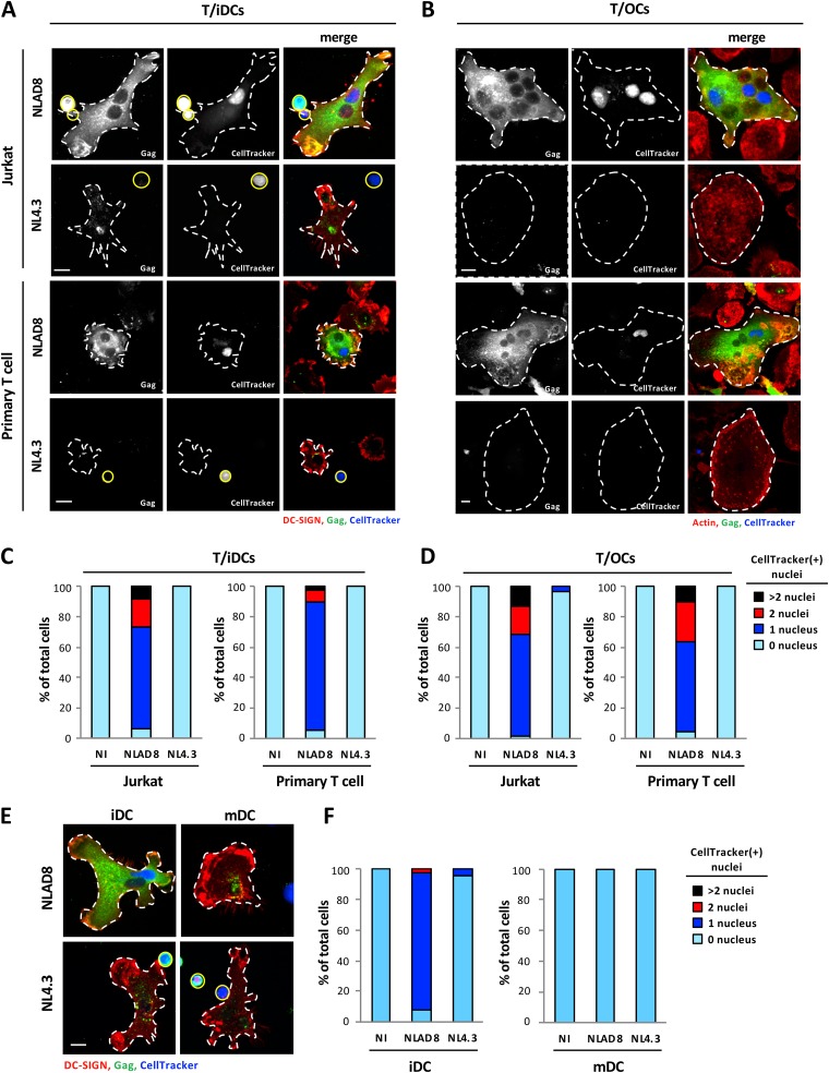
FIG 2.
Viral transfer to immature DCs and OCs by cell fusion with infected T cells. (A and C) NLAD8- or NL4.3-infected Jurkat (A, upper images; C, left panel) or primary CD4+ T (A, lower images; C, right panel) cells prelabeled with CellTracker were cocultured for 6 h with iDCs. Cells were then stained with anti-Gag and anti-DC-SIGN and analyzed by confocal microscopy (scale bar, 10 μm) (A). T cells and iDCs are indicated by plain yellow and dashed white lines, respectively. (C) The number of nuclei per DC-SIGN+ cell was determined from images of at least 50 cells. Results are expressed as the percentages of cells with 0, 1, 2, or more than 2 CellTracker-positive [CellTracker(+)] nuclei. (B and D) NLAD8- or NL4.3-infected Jurkat (B, upper images; D, left panel) or primary CD4+ T (B, lower images; D, right panel) cells prelabeled with CellTracker were cocultured with OCs for 6 h. After elimination of T cells, OCs were stained with anti-Gag and phalloidin (actin) and analyzed by confocal microscopy (scale bar, 10 μm) (B). OCs are indicated by dashed white lines. (D) The number of nuclei per cell was determined from images of at least 50 cells. Results are expressed as the percentages of cells with 0, 1, 2, or more than 2 CellTracker(+) nuclei. (E and F) NLAD8- or NL4.3-infected Jurkat cells prelabeled with CellTracker were cocultured for 6 h with iDCs (E, left images; F, left panel) or mDCs (E, right images; F, right panel). Cells were then stained with anti-Gag and anti-DC-SIGN and analyzed by confocal microscopy (scale bar,10 μm) (E). T cells and DCs are indicated by plain yellow and dashed white lines, respectively. (F) The number of nuclei per DC-SIGN+ cell was determined from images of at least 50 cells. Results are expressed as the percentages of cells with 0, 1, 2, or more than 2 CellTracker(+) nuclei. The results shown are representative of 3 independent experiments performed with DCs and OCs from 3 different donors. NI, noninfected Jurkat or primary CD4+ T cells were cocultured with OCs or DCs.

