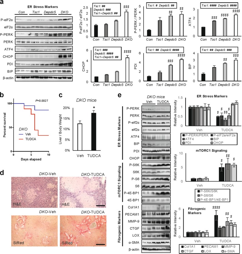
Fig. 5. Relieving ER stress unexpectedly aggravated DKO liver pathologies.
Mouse cohort described in Fig. 2 was subjected to immunoblotting a. Six-week-old DKO mice were injected daily with vehicle (Veh) or 500 mg/Kg TUDCA for 10 days (b–e; n ≥ 9). For drug treatment experiments, mice were gender-matched with both males and females. a ER stress signaling markers were examined from the indicated liver lysates through immunoblotting (left panels) and quantification (right panels). b Mouse survival was monitored throughout the course of the experiment. The P value was calculated through a log-rank test. c Liver/body weight ratio was measured at the experimental endpoint. d Liver sections were analyzed through H&E and SiRed staining. e Liver lysates were subjected to immunoblotting (left panels) and quantification (right panels) to examine ER stress signaling (top), mTORC1 signaling (middle) and fibrogenic markers (bottom). Data are presented as mean ± SEM (n ≥ 3) or actual values b. *P < 0.05 (Student’s t-test). Effects of Tsc1 and Depdc5 mutations and their interaction (Tsc1 × Depdc5) were assessed through two-way ANOVA (#P < 0.05; ##P < 0.01; ###P < 0.001; ####P < 0.0001), and statistical significance between Con and indicated groups were assessed through Tukey’s multiple comparison test ($$P < 0.01; $$$P < 0.001; $$$$P < 0.0001). For western blot quantification, the Holm-Šídák method was used to compare groups ($P < 0.05; $$P < 0.01; $$$$P < 0.0001). Scale bars, 200 µm

