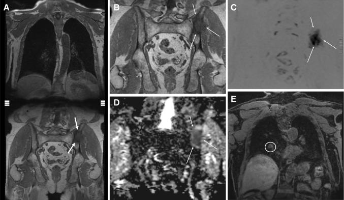
Fig. 1.
73-year-old man with a Gleason 4 + 3 PCa pT3 with PSA of 18.2 ng/ml. A whole-body MR examination was performed. Coronal whole body T1-weighted image (a) shows a left iliac bone metastasis (b; magnification; arrows). The bone metastasis (arrows) are evident on whole-body DWI images (c, d). e In the right lung, para-hilar, a 15 mm metastasis was observed (circle)

