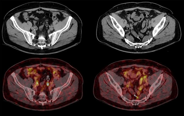
Fig. 3.
72-year-old man with biopsy proven PCa Gleason 4 + 3 pT2c and PSA of 6.7 ng/ml. Preoperative staging with imaging revealed two small lymph nodes in the pelvis, along the left common iliac artery (white arrow) and in the presacral fat (white arrowhead). On CT scan they both have a size of 5 mm which is below the morphological threshold to classify them as suspicious, but on 68Ga PSMA PET-CT they both showed highly avid tracer uptake, indicative of lymph node metastases. Pelvic lymph node dissection was performed and both lymph nodes proved to be malignant. Preoperative staging with 68Ga PSMA PET-CT thus appears to allow for more complete and accurate primary staging of PCa patients compared to standard routine imaging but its role in routine clinical practice is yet to be defined

