Fig. 2.
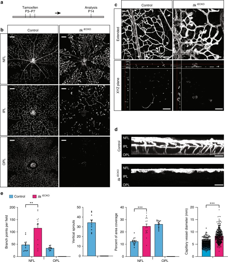
ILK is indispensable for plexus formation in the deeper retina. a IlkiECKO mutants and littermate controls received intraperitoneal tamoxifen injections every other day from P3–7 and were analyzed at P14. b Isolectin B4-stained IlkiECKO mutants and littermate control retinas. Optical sections of z-stacked confocal images were divided to represent the nerve fiber layer (NFL), inner plexiform layer (IPL), and outer plexiform layer (OPL). Note strong reduction of the IlkiECKO IPL and OPL vasculature. Scale bar, 200 μm. c, d, XY-extended, ZY-extended, and XYZ plane view high magnification images of control and IlkiECKO retinas stained with isolectin B4. Vertical sprouting is impaired and abnormal, bulging endothelial structures decorate dilated vessels in IlkiECKO retinas. Scale bar, 100 μm (top) and 50 μm (bottom) in (c), 25 µm in (d). e Quantification of branch points, vascular area coverage, vertical sprouts and capillary vessel diameter, as shown in (b) and (c). Error bars, s.e.m. p values (***p < 0.001, **p < 0.01), Student’s t test (n = 6 retinas/group). Source data are provided as a Source Data file

