Fig. 5.
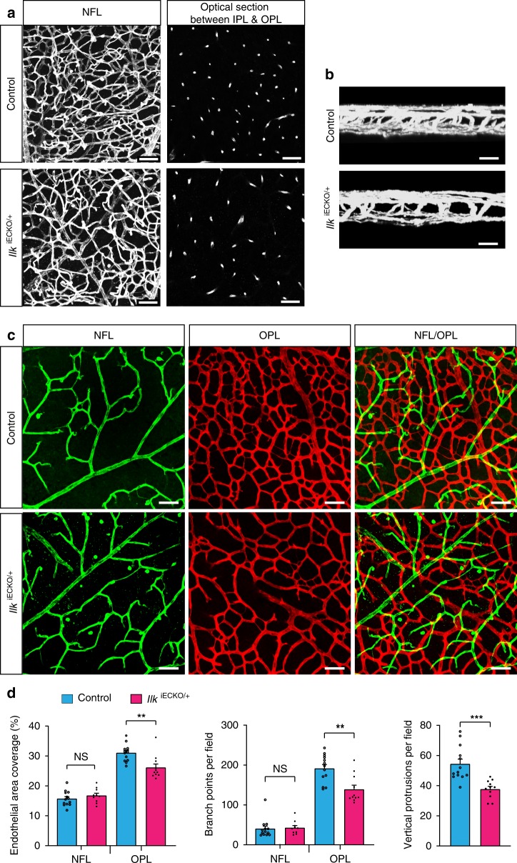
Vascular defects in IlkiECKO/+ heterozygotes. a, b Extended focus (a:XY; b:XZ) and optical section images (right panels in a) of isolectin B4-stained P17 control and IlkiECKO/+ retinal wholemounts. Vessel density and vertical sprouts between IPL and OPL are reduced in IlkiECKO/+. Scale bar, 50 μm in (a) and 25 μm in (b). c, d Images of isolectin B4-stained control and IlkiECKO/+ retinal vessels with pseudo-coloring of the NFL (green) and OPL (red) vasculature (c). Scale bar, 50 μm. IlkiECKO/+ OPL EC area, branch points and vertical sprouts are reduced, whereas NFL vessels are comparable to control samples (d). Error bars, s.e.m. p values (***p < 0.001, **p < 0.01, ns not significant), Student’s t test (n = 3 retinas/group). Source data are provided as a Source Data file

