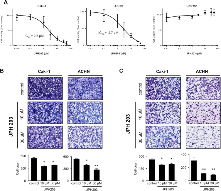
Figure 5.
Effects of JPH203 on the viability, migration and invasion activities of RCC cells. (A) Caki-1, ACHN and HEK293 cells were treated with various concentrations (0.01, 0.1, 1, 3, 10, 30, and 50 μM) of JPH203 for 96 h. The cell viabilities were determined by WST-8 assays, and viability curves are presented as a percentage of the control value, which was obtained from cells treated with DMSO (0.5%). Data are expressed as means with S.E.M. The IC50 values for each cell line were calculated using GraphPad Prism 7 J. The experiments were repeated three times, each performed in triplicate. (B) Transwell chamber migration assays and (C) matrigel invasion assays were used to evaluate the migratory and invasive capabilities of RCC cells following treatment with JPH203 (10 or 30 µM) or DMSO (0.5%) for 48 h. The numbers of cells in five random microscopic fields were counted. Representative images from three independent experiments are shown. Each bar represents the mean with S.E.M. *P < 0.05; **P < 0.01 (unpaired Student’s t-test).

