Fig. 4.
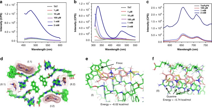
ThT/TmPyP4 binding to Fmoc-G-PNA tetramer. a Fluorescence emission spectra of 20 μM ThT with various concentrations of the Fmoc-G-PNA conjugate, λex = 440 nm. b Fluorescence emission spectra of the Fmoc-G-PNA tetramer in the presence of 20 µM ThT, λex = 290 nm. c Fluorescence emission spectra of 10 μM TmPyP4 with various concentrations of the Fmoc-G-PNA conjugate, λex = 433 nm. d–f Molecular docking of ThT to the Fmoc-G-PNA tetramer. d Binding sites of the first 30 energy-optimized docking patterns. The conformations are clustered into two groups: I.1 and I.2 are equivalent due to the crystal symmetry, as are II.1 and II.2. e, f Interactions mediating the molecule binding in e the first and f the second conformation clusters

