Figure 3.
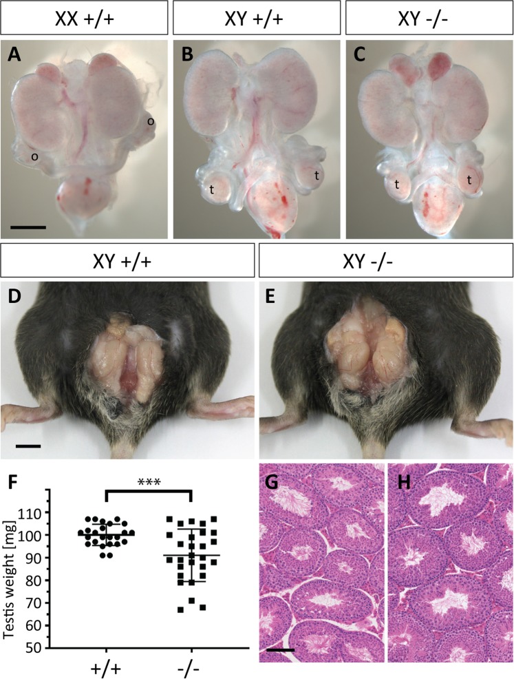
Fetal and adult reproductive structures are overtly normal in Adamts16-deficient mice. (A–C) At 16.5 dpc, the fetal testes (t) have descended to a position adjacent to the bladder in wild-type (B) and Adamts16 −/− mutant males (C); this is in contrast to the para-renal position of the ovaries (o) in wild-type females at the same stage (A). Scale bar = 0.5 mm; (D,E) Testes in adult mice have descended in to the scrotum in wild-type (D) and Adamts16 −/− mutant males (E); scale bar = 0.5 cm; (F) The average weight of the adult testis is reduced in the Adamts16 −/− mutant males (n = 28) compared to wild-type controls (n = 24); ***p ≤ 0.01 (student’s t-test); (G,H) Testicular histology of H&E-stained section appears unaltered in mutant males (H) when compared to wild-type controls (G). Scale bar = 100 μm.

