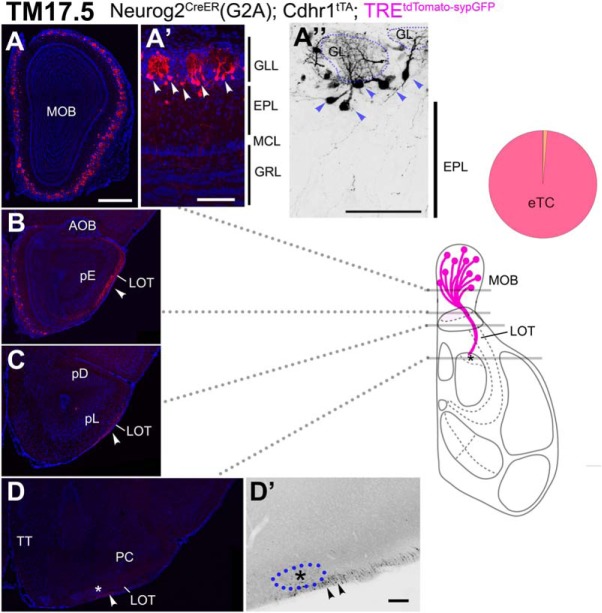
Figure 10.
Birthdate tagging at TM17.5. A–D, Coronal brain sections prepared from a P21 Neurog2CreER(G2A); Cdhr1tTA; TREtdTomato-sypGFP (A–D) mouse that was given TM at E17.5. Images for tdTomato reporter and DAPI. D’, Black and white high contrast image converted from the tdTomato reporter image. A’, A”, High magnifications of the MOB, of which layer positions are indicated on the right-hand end. Arrowheads (A’, A”) indicate cell bodies of birthdate-tagged eTCs that barely have basal dendrites. The glomerulus (GL) is encircled by a dotted line (A”). A small number of TM17.5 axons project in the ventral surface of the LOT (arrowheads in B–D). Asterisks (D, D’) show the anterolateral edge of the OT. The cell cluster encircled by the blue dotted line (D’) is not penetrated by TM17.5 axons (arrowheads). The brain illustration summarizes TM17.5 axon projections and indicates the levels at which individual sections were prepared. The pie chart shows the proportion of neuron subtypes tagged at this TM stage in this reporter line. Representative images from seven mice. Scale bars = 100 μm (A’, A”, D’) and 500 μm (A–D). GRL: granule cell layer, pD: pars dorsalis, pL: pars lateralis, TT: tenia tecta.

