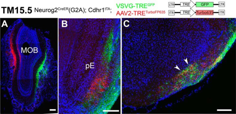
Figure 13.

Topographic and non-topographic maps formed by TM15.5 axons. A–C, Coronal sections prepared from a Neurog2CreER(G2A); Cdhr1tTA mouse that was given TM15.5 and then injected with AAV2-TRETurboFP635 and VSVG-TREGFP viruses in the MOB. Medial is to the left and dorsal is to the top. The eTCs on the medial and lateral sides of the MOB are specifically labeled with red and green reporter proteins, respectively (A). The axons make topographic projections to the pE (B) but not the aiCAP (arrowheads in C). Representative images from four mice with successful focal labeling. Scale bar = 200 μm.
