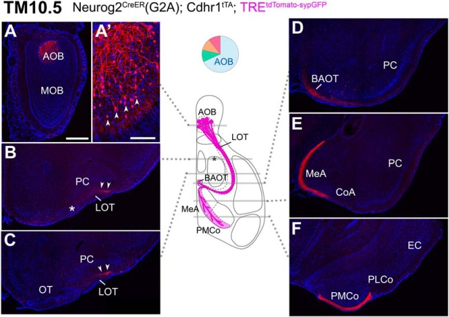
Figure 4.
Birthdate tagging at TM10.5. A–F, Coronal brain sections prepared from a P22 Neurog2CreER(G2A); Cdhr1tTA; TREtdTomato-sypGFP mouse that was given TM injection at E10.5. Images for tdTomato reporter (red) and DAPI (blue). Medial is to the left and dorsal is to the top. The TM10.5 tagging selectively labels AOB projection neurons (A, arrowheads in A’). Their axons run in the deep dorsal side of the LOT (arrowheads in B, C) and project to the AOB-specific targets (D–F). Asterisks indicate the position of the anterolateral edge of the OT. Brain illustration in the middle summarizes TM10.5 axon projections and indicates the levels at which individual sections were prepared. The pie chart shows the proportion of neuron subtypes tagged at this TM stage with this reporter. Representative images from five mice. Scale bar = 100 μm (A’), 500 μm (A–F). BAOT: bed nuclei of the accessory olfactory tract, CoA: cortical amygdala, EC: entorhinal cortex, MeA: medial amygdala, PLCo: posterolateral cortical amygdala, PMCo: posteromedial cortical amygdala.

