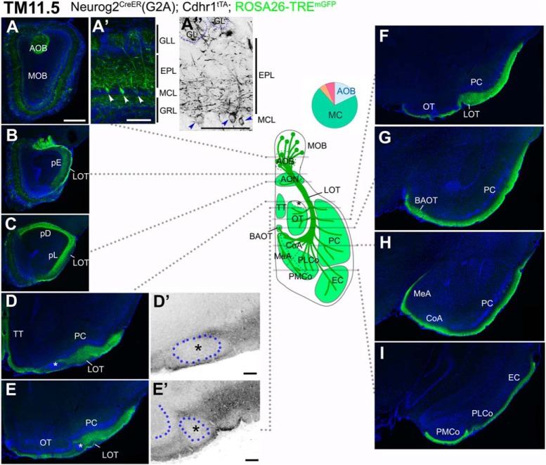
Figure 5.
Birthdate tagging at TM11.5. A–I, Coronal brain sections prepared from a P21 Neurog2CreER(G2A); Cdhr1tTA; ROSA26-TREmGFP mouse that was given TM at E11.5. Images for mGFP reporter (green) and DAPI (blue). A”, D’, E’, Black and white high contrast image converted from the mGFP image. A’, A”, Higher magnifications of the MOB, of which layer positions are indicated on the right-hand end. Arrowheads (A’, A”) indicate cell bodies of birthdate-tagged MCs. Note that the basal dendrites in the lower part of the EPL are labeled with the reporter. The glomerulus (GL) is encircled by a dotted line (A”). The TM11.5 axons project diffusely to all the MOB and AOB targets (B–I) except the small domain in the anterolateral edge of the OT (asterisks in D, D’, E, E’). D’, E’, Position of a cell cluster is encircled by the blue dotted line and marked with the asterisk. The curved dotted line on the left side of the cell cluster in E’ depicts the lateral hook of the OT cell layer. The labeling of the LOT axons looks less intense possibly because the axons are highly myelinated (Inaki et al., 2004). The brain illustration in the middle summarizes TM11.5 axon projections and indicates the levels at which individual sections were prepared. A pie chart shows the proportion of neuron subtypes tagged at this TM stage with this reporter. Representative images from six mice. Scale bars = 100 μm (A’, A”, D’, E’) and 500 μm (A–I). BAOT: bed nuclei of the accessory olfactory tract, CoA: cortical amygdala, EC: entorhinal cortex, GRL: granule cell layer, MeA: medial amygdala, pD: pars dorsalis, pL: pars lateralis, PLCo: posterolateral cortical amygdala, PMCo: posteromedial cortical amygdala, TT: tenia tecta.

