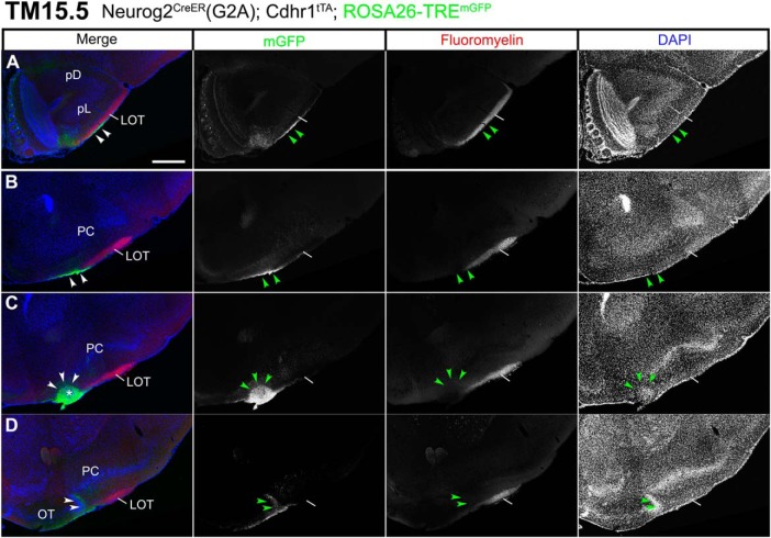
Figure 9.
Segregated projections of TM15.5 axons to the target cell cluster in the anterolateral OT. A–D, Coronal brain sections prepared from a P22 Neurog2CreER(G2A); Cdhr1tTA; ROSA26-TREmGFP mouse that was given TM at E15.5. Images for mGFP reporter (green), FluoroMyelin (red) and DAPI (blue). FluoroMyelin marks myelinated OB axons running through the LOT. The TM15.5 axons (arrowheads) ventrally segregate from other OB axons in the LOT (A, B) and terminate at a small cell cluster in the anterolateral edge of the OT (arrowheads and asterisk in C). In a posterior section (D), the cell cluster fuses with the lateral hook of the OT dense cell layer (arrowheads). Representative images from five mice. Scale bars = 500 μm (A–D). pD: pars dorsalis, pL: pars lateralis.

