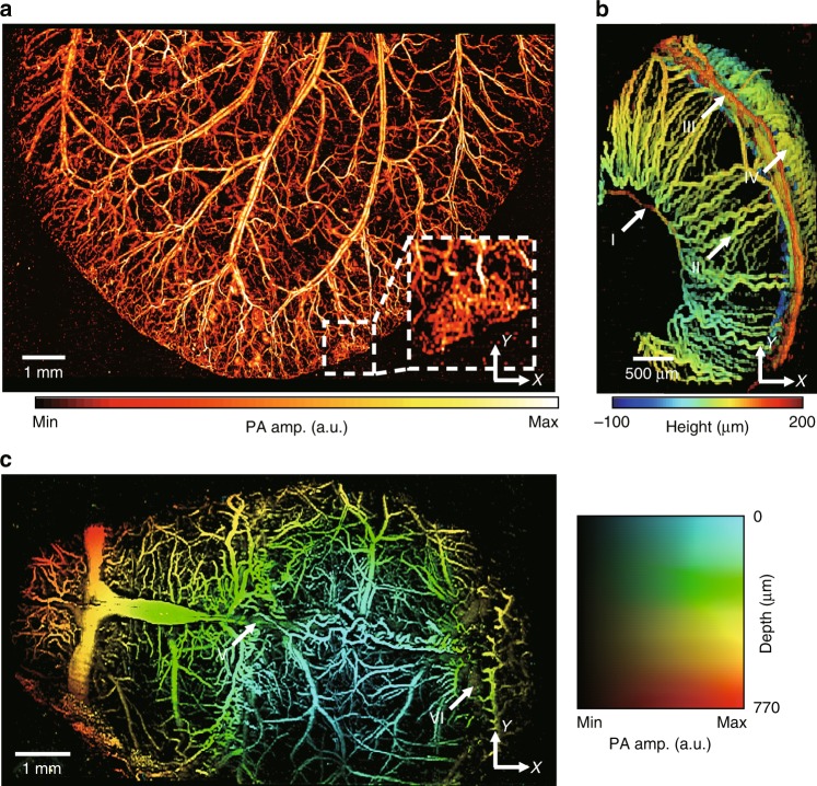
Fig. 2. Photoacoustic (PA) images of microvasculatures in small animals in vivo.
a Wide-FOV PA MAP image of a mouse ear. The region including a capillary bed is outlined by the white dashed box. b Depth-encoded PA image of a mouse eye. Circulus arteriosus major (I), iris (II), circulus arteriosus minor (III) and choroid (IV) blood vessels are highlighted by the white arrows. c Wide FOV PA MAP image of a mouse brain with color-encoded depths and amplitudes. Superior sagittal sinus (V) and transverse sinus (VI) are highlighted by the white arrows. FOV, field of view; MAP, maximum amplitude projection.

