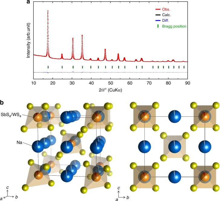
Fig. 2.
Crystal structure of Na2.88Sb0.88W0.12S4 heated at 275 °C for 12 h. a Rietveld refinement profiles of X-ray powder diffraction data for Na2.88Sb0.88W0.12S4. Red dots and black lines denote the observed and calculated XRD patterns, respectively. The green sticks mark the position of the reflections for Na2.88Sb0.88W0.12S4. The difference between the observed and calculated patterns is indicated by the blue line. b Crystal structure of cubic Na2.88Sb0.88W0.12S4 with the unit cell outlined. The Na, Sb, W, and S sites are represented by blue, orange, gray, and yellow balls, respectively. Na is linearly arranged, and distortion of the Sb4/WS4 tetrahedra is very small compared to the tetragonal structure of Na3SbS4 (Supplementary Fig. 3)

