Figure 6.
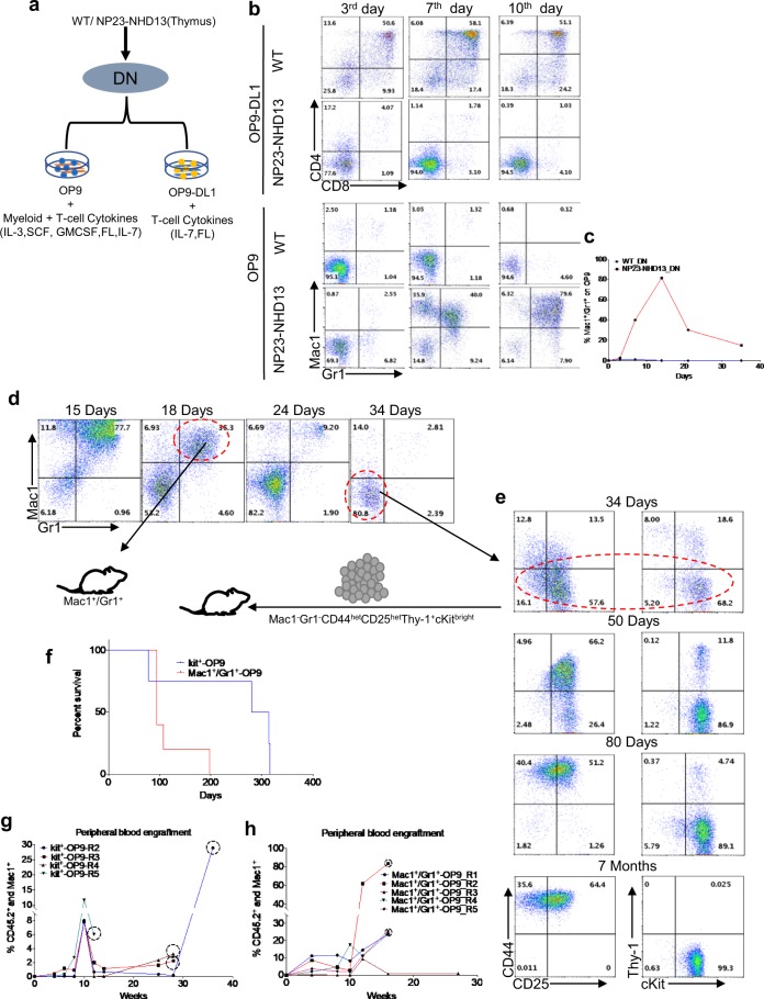
In vitro differentiation of NP23-NHD13 DN thymocytes. (a) In vitro co-culture of DN cells from WT and NP23-NHD13 mice on OP9 and OP9-DL1 feeder layers with the indicated cytokines; DN cells were also Mac1−(1 × 10⁵ cells were plated). (b) Differentiation of DN cells on OP9 and OP9-DL1 at indicated timepoints. (c) graph represents Mac1+/Gr1 + percentage of WT and NP23-NHD13 DN cells on OP9. (d,e) Long term co-culture of DN cells from NP23-NHD13 mice on OP9. Mac1+/Gr1+ and Mac1−Gr1−CD44hetCD25hetThy-1+cKitbright (designated Kit + -OP9) cells (1 × 10⁴ cells/mouse) were transplanted at 18 or 34 days of culture, respectively. (f) Survival of Mac1+/Gr1+ and Kit + -OP9 recipients. Data are analyzed by Log-rank (Mantel-Cox) test), P = 0.07. Engraftment of donor myeloid cells (CD45.2+/Mac1+) is shown in both (g) Kit + -OP9 and (h) Mac1+/Gr1+ recipients (dotted circle indicates engraftment data obtained from BM).

