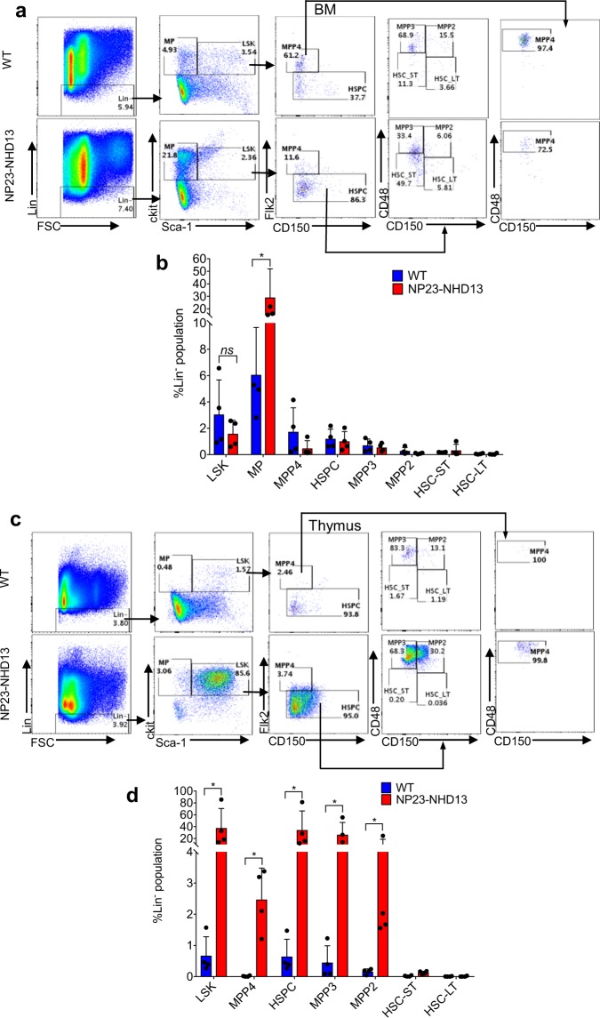
Figure 8.
Thymi from non-leukemic NP23-NHD13 mice show a markedly expanded LSK compartment. (a) Flow cytometry of WT and NP23-NHD13 BM using Flk2, CD48, and CD150 staining to fractionate LSK cells (b) Quantification of LSK cells and LSK subpopulations in BM nsP = 0.17(LSK). (c) Flow cytometry of WT and NP23-NHD13 thymus (d) Quantification of LSK cells and LSK subpopulations in thymus. Data are expressed as means ± SD, n = 4 mice per group, *P < 0.05, by Mann Whitney test.

