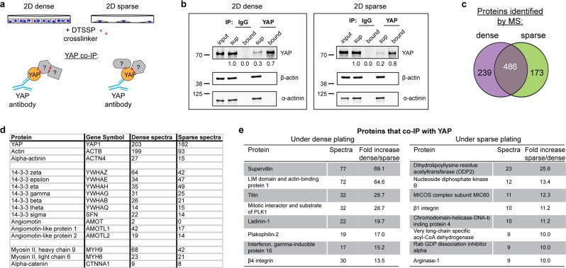
Figure 3.
Protein that co-IP with YAP under activating or inactivating conditions. (a) Schematic of YAP co-IP from cells crosslinked with 5 mM DTSSP while sparsely or densely plated on 2D tissue culture plastic. (b) Western blot analysis of YAP co-IP from cells crosslinked with 5 mM DTSSP while sparsely or densely plated on 2D tissue culture plastic. Bound is 5x load with respect to input. Fluorescence intensity quantification of YAP indicated below each band. Images cropped from the same blot using different fluorescent intensities, stripped, and re-probed. Full-length blot included in Supplementary Fig. 1. (c) Venn diagram showing overlap of proteins and number of YAP and actin spectra identified by UPLC-MS/MS to co-IP with YAP under dense or sparse conditions. (d) Spectra of YAP, actin, α-actinin, and proteins previously identified as YAP interactors. (e) Proteins that differentially co-IP with YAP under sparse or dense conditions.

